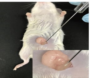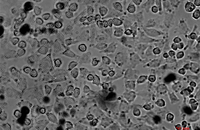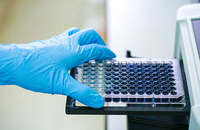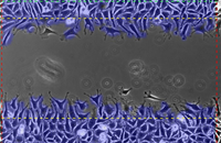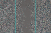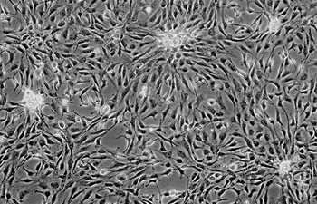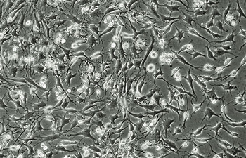
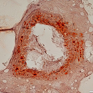

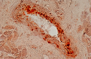
Various types of cells isolation and culture
The importance of performing primary culture of animal cells to evaluate the effectiveness and toxicity of new drugs, vaccines, biological drugs as well as reproductive technology has been proven. In Histogenotech, isolation of different types of cells from various human and animal tissues, including skin, fat, neurosphere, cornea, etc., can be performed using specific enzymes and mechanical methods.

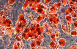
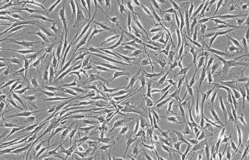
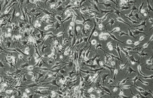
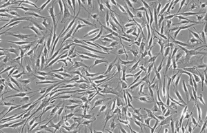
Differentiation of cells into different cell lines
Cultivation of pluripotent stem cells (PSCs) and their differentiation into different cell types has become an important factor in medicine and drug development. Histogenotech, with be beneficial of the latest technical knowledge and the best brand of cell culture media and other requirements, has been able to run the process of differentiation PSCs into different cell lines of nerve, cartilage, bone, fat, cardiomyocytes. Also, confirming of differentiation will perform using flow cytometric methods and immunocytochemistry.
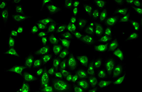
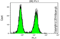
Confirmation of cell nature by flow cytometry and ICC
Demonstration of induction of stem cell differentiation into varying cell lines can be done by identifying the specific antigens of cell by immunofluorescence method. Histogenotech has been able to confirm cells differentiated into different cell types in different signaling pathways by using specific antibodies of different cell lines.
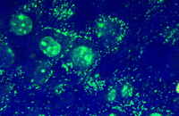
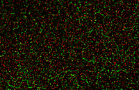
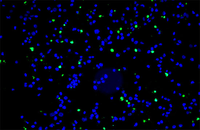
Examination of Live and Dead Cells
Acridine orange and propidium iodide (PI) are nucleic acid-binding dyes used to measure cell viability. By acridine orange staining, all nucleated cells are stained to produce a green fluorescence. While PI dye only enters cells with damaged membranes and turns dying or dead nuclear cells red. In addition, other membrane viable dyes such as ethidium bromide, 7AAD, SYTOX, DRAQ5 may be used instead of PI. In Histogenotech, cell viability is assessed by fluorescent image analysis and the ratio of live to dead cell can calculate.
Investigation of cancer cell migration using Transwell
One of the most important characteristics of malignant tumor cells is their ability to invade and migrate. Studies on cell migration are invaluable for the diagnosis, prognosis, drug development, and treatment of cancer. For this reason, there are several methods for examining this process in Histogenotech. The most common methods in Histogenotech are transwell migration assay, scratch wound assay, and molecular measurement of matrix metalloproteinase (MMP) as the major enzyme in extracellular matrix degradation.
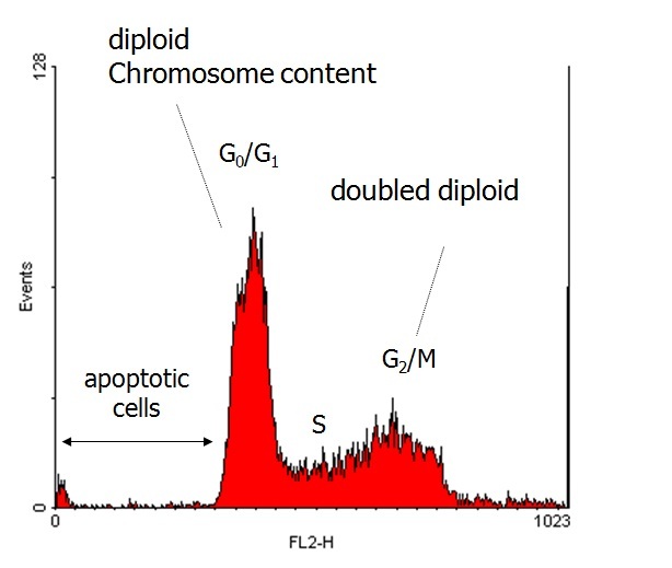
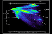
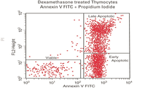
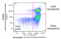
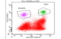
Evaluation of cell apoptosis by flow cytometry
Determining apoptosis, or programmed cell death, is important in the different animal studies and in many diseases, including cancer. At the Histogenotech Research Center, cell apoptosis is performed by a variety of methods, including cell staining with annexin PI, anxin V, tunnel and fluorescent. By histogram analysis, the results of these techniques can be reported for cell necrosis.
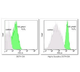
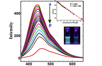
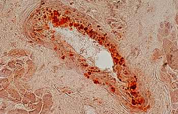
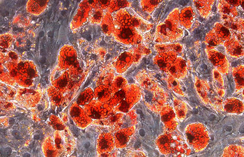
Investigation of cell antioxidants by spectrofluorometric
In Histogenotech, the level of reactive oxygen species (ROS) in frozen tissue samples can be measured based on the conversion of dichlorofluorescein acetate (DCFDA) using fluorimetry techniques. For this reason, after staining the tissue with DCFDA in the dark place, the absorbance of the samples at 488nm and 593nm is measure using a fluorimeter, which finally determines the ROS level based on the H2O2 standard curve.
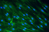
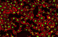
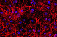
Check the placement of the cell on the scaffold
The most important part in the performance of various synthetic scaffolds is the study of cell adhesion on the scaffold. In the Histogenotech, there are various methods for examining cell adhesion that are used depending on the type and material of the scaffold. One of the most common methods of staining is cytoskeleton analysis with rhodamine / phalloidin and nucleus arrangement with DAPI. In this method, the extent of cytoskeleton and nucleus binding is assessed by fluorescent microscopy imaging.
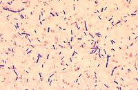
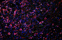
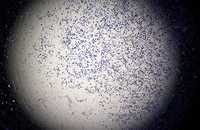
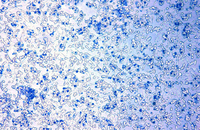
Investigation of cellular contamination
One of the major problems of students is the presence of microbial contamination, especially Mycoplasma contamination in cell culture media. Histogenotech can detect a variety of microbial contamination by staining cells with Hoechst fluorescent dye