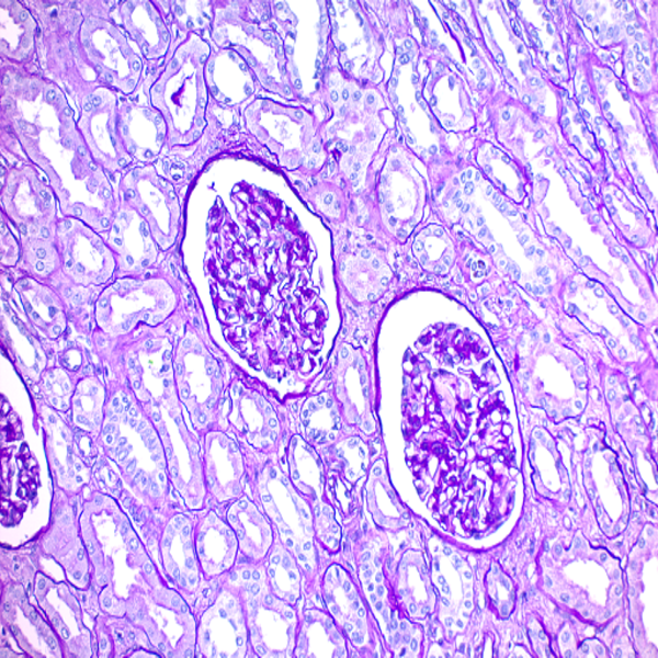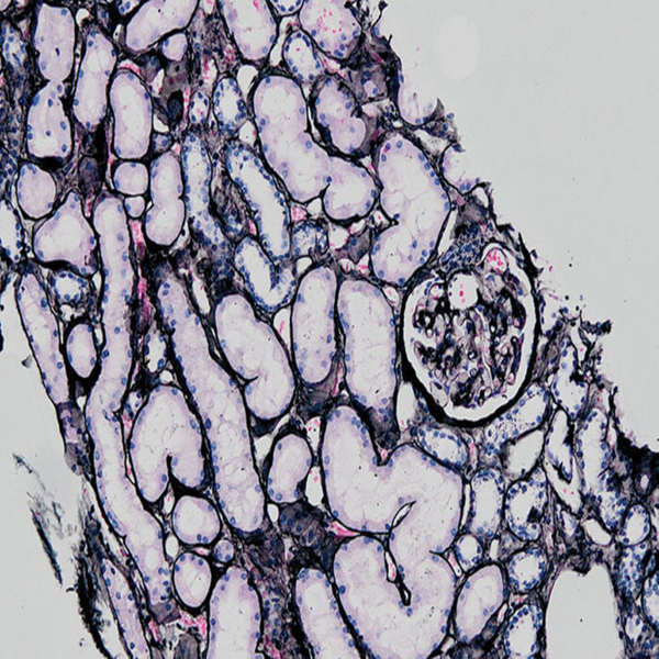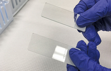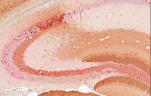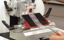
Preparation of paraffin and frozen tissue sections
Tissues can be store for long periods of time through paraffin molding and freezing. In Histogenotech samples are molded after tissue dehydration by paraffin dispenser. Also microtome cutting machine is use for tissue preparation for various staining.
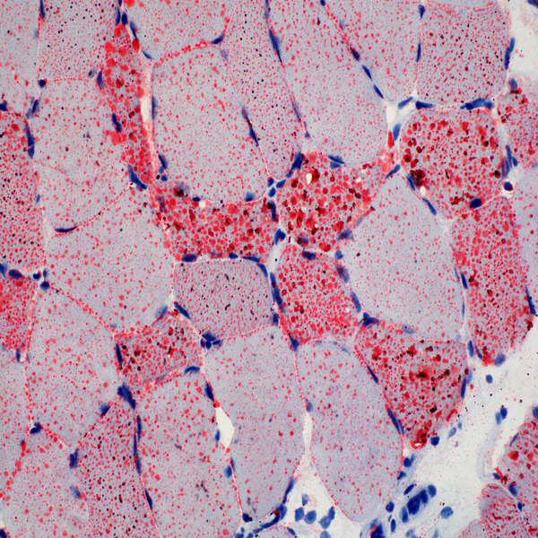
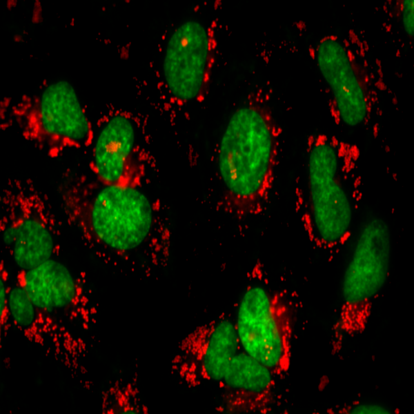
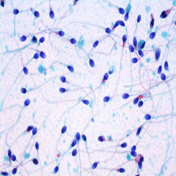
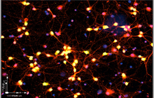
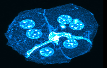
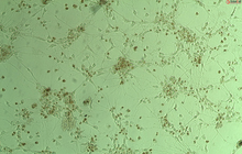
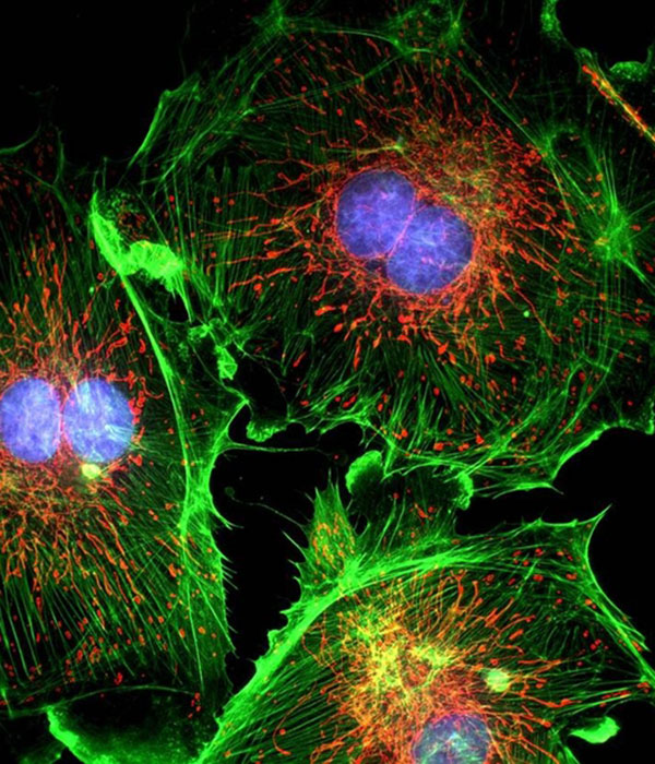
Immunocytochemical technique
Immunocytochemistry is a common technique to detect specific proteins or antigens in cells using a variety of antibodies. Histogenotech is ready to provide immunocytochemical services for researchers due to stored antibody bank and existence of light and fluorescent microscopes in laboratory.
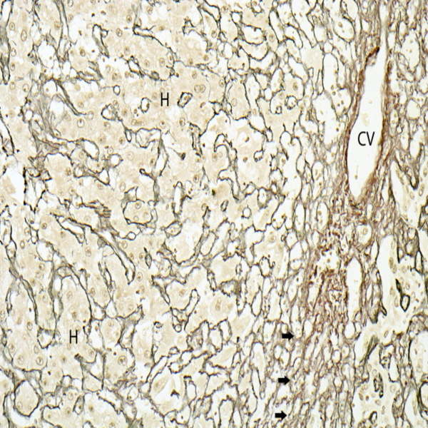
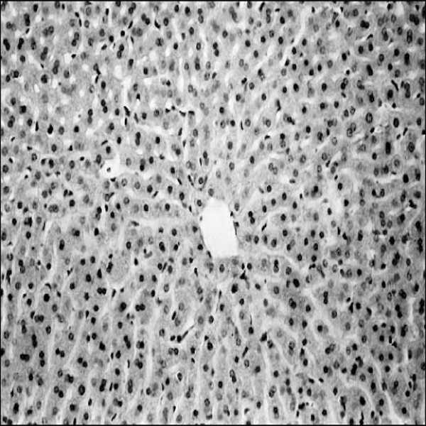
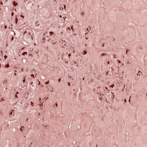
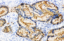
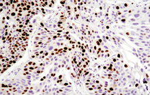
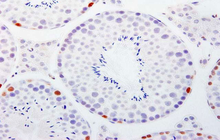
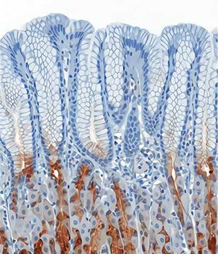
Immunohistochemical technique
This technique is used to study the expression of a variety of proteins in different tissue. Histogenotech and his trained specialist are ready to provide immunohistochemical services for professors, students and researchers with a bank of antibodies and specific histological dyes.
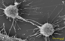
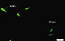
Examination of apoptosis (tunnel)
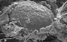
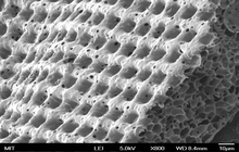
The SEM microscope is perform to study the morphology of nanostructures and to identify compounds as well as to accurately examine the cell surface. To complement its histological and cellular services, Histogenotech has also put on its agenda the evaluation of chemical composition gradients, breakdown stages and the identification of compounds on the surface of samples.
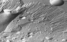
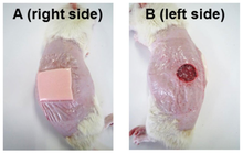
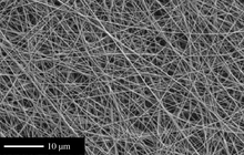
Preparation of tissue scaffolds
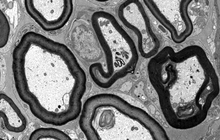
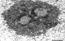
Structural examination of tissues and cells plays a crucial role in advancing new studies at Histogenotech histological unit because in modern research, the morphology of cells and organs is examined by TEM micrograph.
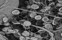
Evaluation of the cells on the scaffold
The SEM microscope is perform to study the morphology of nanostructures and to identify compounds as well as to accurately examine the cell surface. To complement its histological and cellular services, Histogenotech has also put on its agenda the evaluation of chemical composition gradients, breakdown stages and the identification of compounds on the surface of samples.
Preparation of tissue sections for examination with a TEM microscope
Structural examination of tissues and cells plays a crucial role in advancing new studies at Histogenotech histological unit because in modern research, the morphology of cells and organs is examined by TEM micrograph.
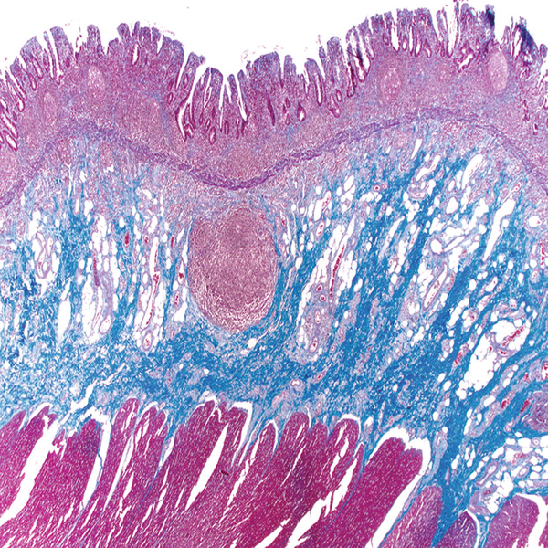
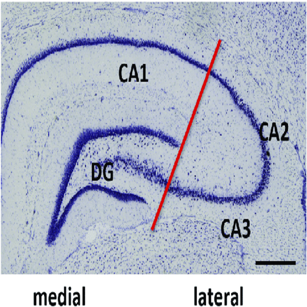
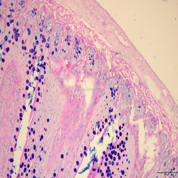
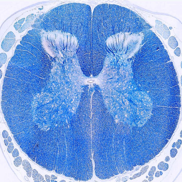
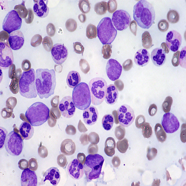
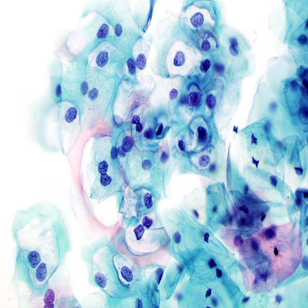
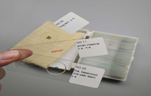
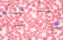
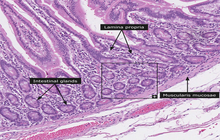
Preparation of training slides
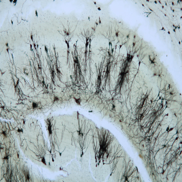
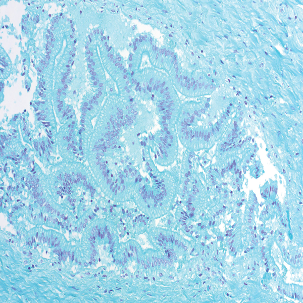
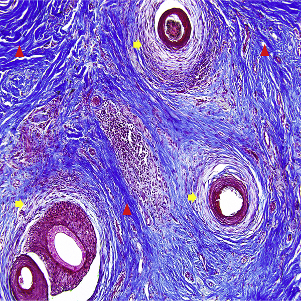
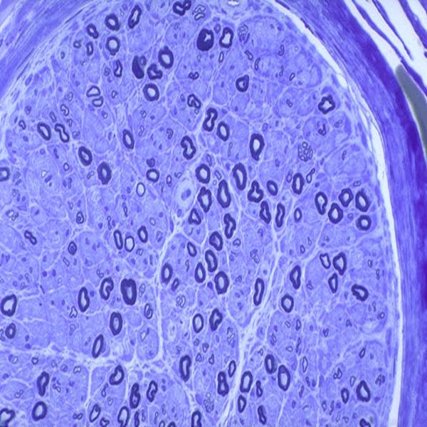
Histogenotech in order to improve the scientific level of students and also to facilitate the educational process for university attends in the fields of histology, embryology, hematology, cellular and molecular, has prepared educational slides from all tissues and organs of the body.