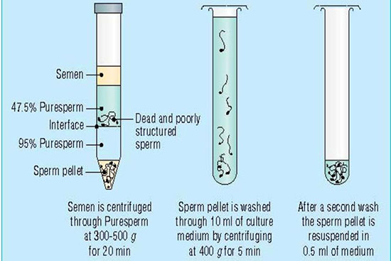
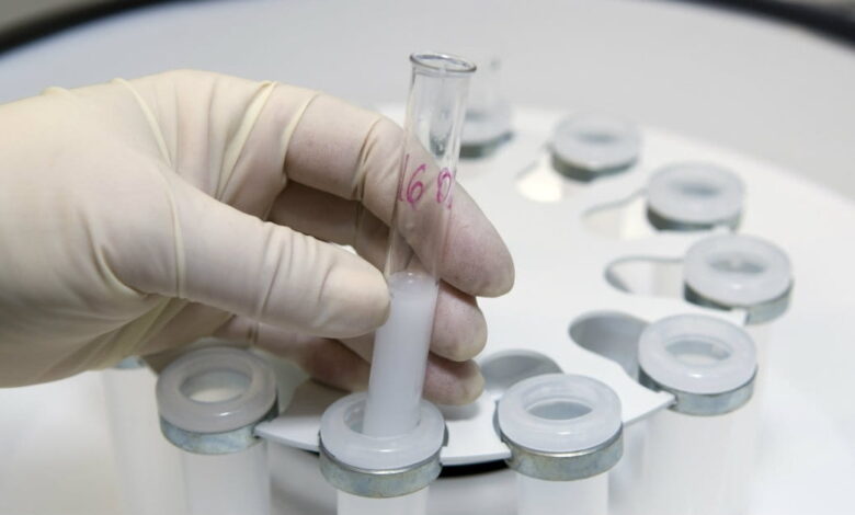
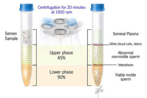
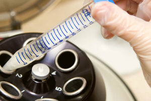
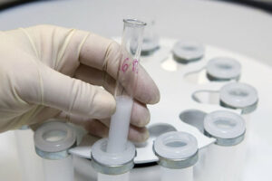
Isolation and washing of sperm
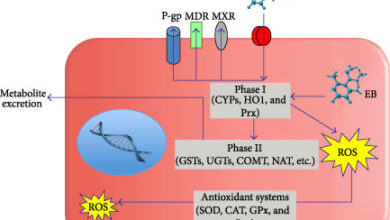
Ideal sperm isolation should be rapid, easy, and cost-effective, to separate motile sperm, not to damage sperm or abnormal changes in isolated sperm cells, remove inactive sperms and other cells including leukocytes, bacteria, toxins or bioactive substances such as decapitation factors or reactive oxygen species (ROS). In the conventional swim-up method, functional sperm can be centrifuged in close cell-to-cell contact with defective sperm or leukocytes. As a result, they cause massive oxidative damage to the plasma membrane of sperm by ROS and consequently sperm function. Thus, other separation methods such as concentration gradient or sperm filtration with Sephadex column and membranes are suggested. These methods are widely used in Histogenotech with the beneficial of experienced specialists.
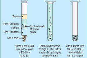
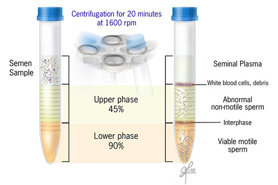
Provide wash culture medium and sperm concentration gradient
Sperm washing is often performed to obtain high-quality specimens (greater sperm count and motility). Different methods of sperm separation can be classified into migration methods, density gradient centrifuges and filtration methods. A prerequisite for all methods of sperm migration and motility. For density gradient centrifuges, isolated culture media such as Ficoll and Percoll have been introduced. Glass wool filtration and sperm filtration with Sephadex columns and membranes are alternative separation techniques. The typical medium available in andrology or embryology laboratories as well as the embryology department of the Histogenotech, Ham F-10 and HTF is enriched with HAS (Human Serum Albumin). These methods are used frequently in the master and PhD thesis of different branches of developmental biology, embryology and dissection sciences,.
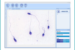
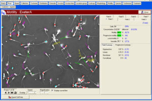
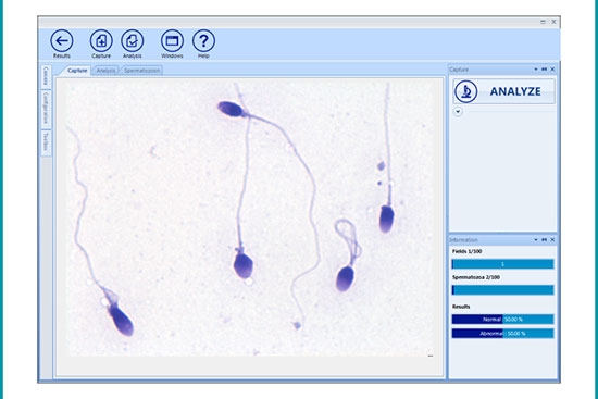
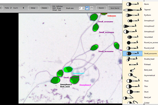
Manual spermogram and CASA
Spermogram is a laboratory analysis of male sperm that is performed to estimate the fertility of sperm. In an effort to make semen quality assessment more targeted and accurate, tools for computer-aided semen analysis (CASA) have been developed. Using CASA, several specific motility parameters can be obtained that describe sperm motions (Progressive, Motile, Sluggish, Immotile) in more detail. Since sperm concentration is a strong predictor of fertility in men, the Histogenotech Reproductive Research Laboratory has developed various research projects on the use of supplements (vitamin C, L-carnitine, pentoxifylline, various herbs) to increase the rate of fertility. The motility and ultimately fertility of adult male mice is maintained in normal sperm culture medium or frozen and thawed sperm.

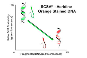
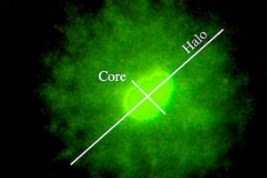
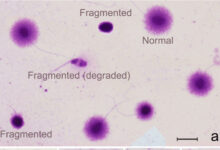
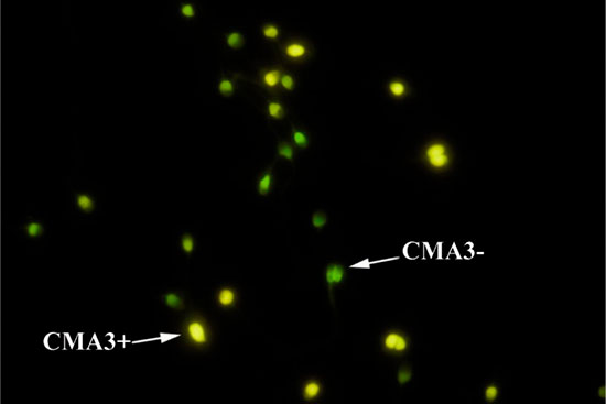
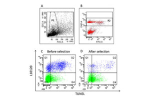
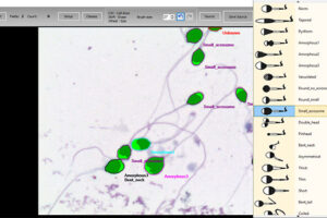
Specialized staining of sperm chromatin
The diagnosis of male infertility is mainly based on the concentration, motility and morphology of semen parameters based on the World Health Organization (WHO). However, none of these parameters can predict a couple’s fertility potential. Sperm Chromatin Structure Test (SCSA) is a flow cytometric test that measures the sensitivity of sperm DNA to the denaturation of in situ acid-induced DNA. DNA fragmentation of sperm, measured by SCSA, is a predictor of successful pregnancy and can be used as a tool for screening, counseling, and treating infertility without cause. Tunnel method for sperm DNA degradation, chromatin staining with chromomycin A3 staining, acridine orange staining, Comet Assay method, measuring DNA degradation by halosperm method are all methods that can be performed in Histogenotech.


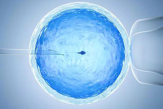
Sperm, egg and embryo manipulation medium
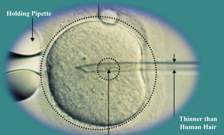
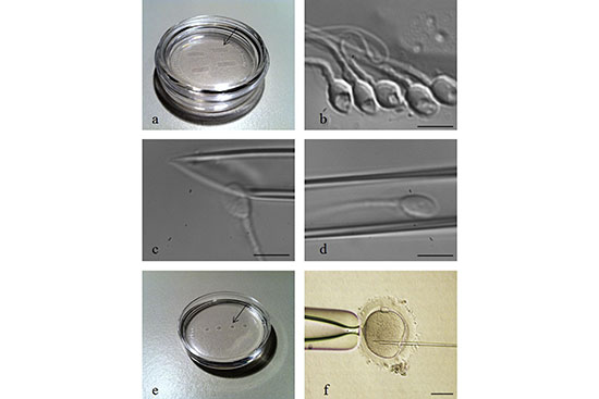
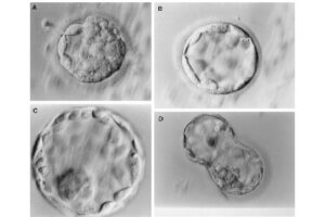
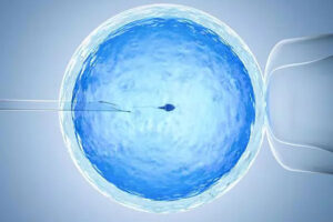
Intracytoplasmic sperm injection (ICSI), in vitro fertilization (IVF), in vitro egg maturation (IVM) and in vitro sperm preparation require different culture media called sperm, egg and embryo manipulation medium. In order to achieve a successful fertility, sperm must maintain their motility and survival in a suitable culture medium in terms of pH, osmolality and sufficient nutrients, and reach the egg to be fertilized. On the other hand, MI or MII oocytes in primary (primary), secondary (preantral), tertiary (graph or antral) follicles must maintain their adult maturity or survival in the laboratory in order to fertilize with quality sperm, quality embryo Create a 2PN in step. In order to increase the fertility rate, a suitable embryo culture medium will enable 2PN embryos to grow into 2 cells, 4 cells, 8 cells, 16 cells, or morula and blastocysts.
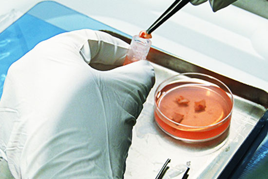
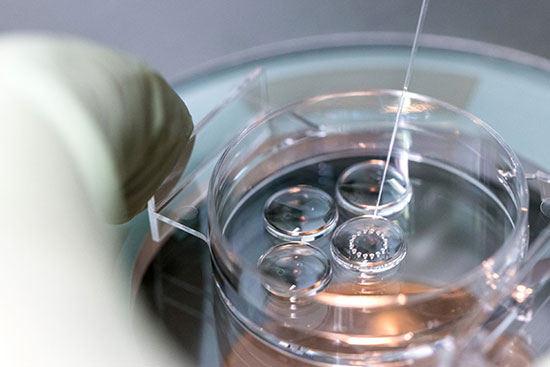
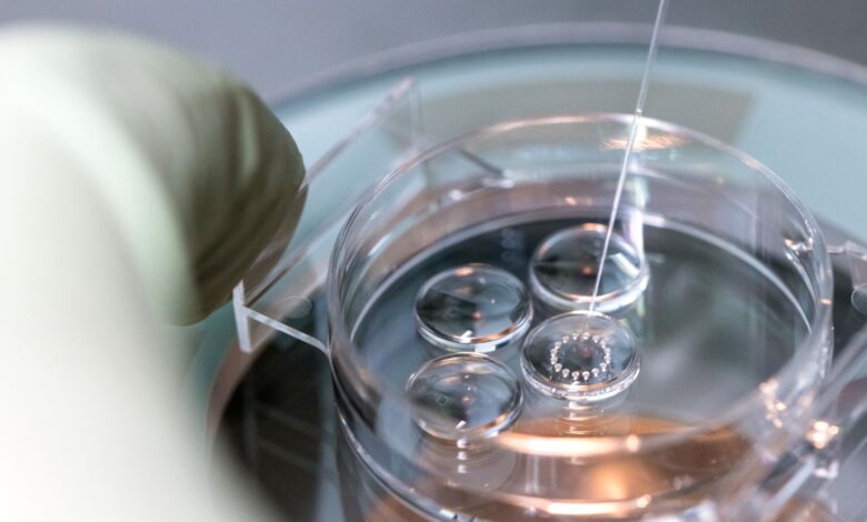
Egg preparation and in vitro fertilization (IVF)
In vitro fertilization is an assisted reproductive technique (ART) commonly referred to as IVF. In vitro fertilization involves the fertilization process, in which an egg cell and a sperm cell combine manually in the laboratory. The resulting blastocyst or morula embryos are transferred into the uterus to continue the developmental process and implant in the body to continue the developmental stages. Other methods of assisted reproduction include the transfer of male germ cells into the fallopian tube (GIFT) and the transfer of embryos at different stages of development into the fallopian tube (ZIFT). In order to perform microinjection processes (ICSI) or in vitro fertilization (IVF) and egg freezing, egg preparation (incubation) is required. Thus, the eggs collected during ovulation or puncture are collected at different stages of nuclear maturation, including GV, GVBD, and MI eggs in a Histogenotech laboratory, and after being placed in a fertilization drop, become a fertilized MII egg or a 2PN embryo.


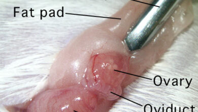
Stimulation of ovulation and separation of ovarian follicles
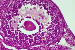
Oocyte pick-up (OPU) or in vitro fertilization (IVM) or in vitro fertilization (IVF) is one of the most common aspects of assisted reproductive technology. Because in vitro fertilization requires the injection of ovulation-stimulating hormones for in vitro fertilization, access to mature eggs, with a limited number and not more than usual, can lead to a successful fertilization. In order to receive more eggs and achieve more embryos for a successful in vitro fertilization, it is necessary to create hormonal conditions appropriate to the sexual cycles of female mice. In this regard, by injecting gonadotropins such as HMG, Letrozole FSH and PMSG, more primitive or primary follicles can be stimulated in the human or mouse body to resume the process of ogenesis and mature. The collected eggs are located inside the follicles with different dimensions and stages of nucleus maturation. After harvesting in the Histogenotech embryology laboratory, they are germinated (removal of cumulus cells around the egg and granulosa cells) and after culture in a suitable medium using oocyte culture, washed sperm are fertilized.